Abstract
Background: Liver cirrhosis frequently precipitates malnutrition due to impaired nutrient intake and metabolic dysregulation, consequently elevating morbidity and mortality. The prompt identification of malnutrition is essential for averting complications.
Objective: This study sought to evaluate the nutritional status of cirrhotic patients employing the Mini Nutritional Assessment (MNA) tool.
Method: This cross-sectional investigation enrolled 330 patients, aged 18 years and above, with a confirmed diagnosis of liver cirrhosis. Individuals with malignancies or advanced hepatic encephalopathy (grades 3 and 4) were excluded. Data collection encompassed anthropometric measurements, including Body Mass Index (BMI), Mid Arm Circumference (MAC), and Triceps Skinfold Thickness (TSF). Nutritional status was evaluated using the MNA scoring system, and disease severity was gauged via the Child-Pugh score. Statistical analyses were conducted to explore associations between malnutrition and various clinical parameters.
Results: The mean age of participants was 54.73 ± 11.79 years, with a mean BMI of 22.41 ± 2.76 and a mean MNA score of 19.03 ± 4.43. Malnutrition demonstrated a higher prevalence among older patients (p=0.001), those with hepatitis B etiology (p=0.002), individuals with advanced cirrhosis (Child-Pugh B and C, p=0.000), and those presenting with moderate to severe ascites (p=0.000). Logistic regression analysis identified ascites and hepatic encephalopathy as significant predictors of malnutrition.
Conclusion: Malnutrition is exceptionally common in the cirrhotic population, particularly among those with advanced disease and accompanying complications like ascites. The integration of routine nutritional screening using the MNA can facilitate early detection and management of malnutrition, thereby potentially enhancing patient outcomes.
Keywords: Liver Cirrhosis, Malnutrition, Mini Nutritional Assessment, Nutritional Status, Ascites, Hepatic Encephalopathy, Child-Pugh Score, Cross-Sectional Study.
Introduction
Cirrhosis represents the final common pathway for most chronic liver pathologies, culminating in portal hypertension and end-stage liver disease 1. Its etiology is multifactorial, encompassing chronic hepatitis, alcohol consumption, and various metabolic disorders 2. Globally, chronic liver disease (CLD) is responsible for approximately 2 million deaths annually 3.
Malnutrition is a prevalent complication in individuals with liver cirrhosis, arising from diminished nutrient intake, impaired hepatic metabolism, and reduced intestinal absorption. Its severity tends to escalate with the progression of liver disease 4. It is estimated to affect 65–90% of patients with decompensated cirrhosis 5. Malnutrition significantly exacerbates both mortality and morbidity in CLD patients, with affected individuals demonstrating higher rates of hepatic encephalopathy, infections, variceal hemorrhage, and refractory ascites 6. Furthermore, malnutrition stands as an independent predictor of death 7, 8. Consequently, early screening, assessment, and intervention for malnutrition are paramount, as they aid in preventing complications and enhancing quality of life 9.
Several screening tools are commonly employed for patients at nutritional risk, including the Subjective Global Assessment (SGA), the Malnutrition Universal Screening Tool (MUST), the Nutrition Risk Screening 2002 (NRS-2002), and the Mini Nutritional Assessment (MNA) 10. Numerous studies have validated the reliability and efficacy of the MNA in both cirrhotic patients and the elderly non-cirrhotic population 10, 11, 12, 13. The MNA is a straightforward, non-invasive, economical, and dependable instrument that can be effectively administered by non-dietetic professionals following minimal training 14. Complementary anthropometric assessments in cirrhotic patients include measurements such as body weight, estimated dry weight, height, Body Mass Index (BMI), Mid Arm Circumference (MAC), Triceps Skinfold Thickness (TSF), Mid-Arm Muscle Circumference (MAMC), and Calf Circumference (CC) 15. This study aimed to evaluate the nutritional status of patients with chronic liver disease utilizing the Mini Nutritional Assessment scoring system.
Method
After approval from the institutional ethical review board (IRB No: HMC-QAD-1636, dated 08-12-2023), this cross-sectional study was conducted in the Department of Gastroenterology at Hayatabad Medical Complex, Peshawar, from December 2023 to July 2025. A total of 330 patients were enrolled using a non-probability convenience sampling technique. Written informed consent was secured from all eligible participants prior to enrollment. All patients aged ≥ 18 years with an established diagnosis of liver cirrhosis of any etiology were eligible. Diagnosis was based on clinical criteria (e.g., CLD complications including grade 1 or 2 hepatic encephalopathy, variceal bleeding, ascites), radiological evidence (coarse echotexture on abdominal ultrasound or CT scan), and/or elastographic findings (liver stiffness > 14 kPa) indicative of advanced disease. Patients were excluded if they presented with hepatocellular carcinoma or any other malignancy, heart failure, diabetes mellitus, AIDS, chronic renal failure, grade 3 or 4 hepatic encephalopathy, tuberculosis, or neuropathy. Individuals currently receiving enteral nutritional supplements were also excluded. Strict adherence to these criteria was maintained to mitigate potential confounding variables. Data were recorded on a pre-designed questionnaire. This included biochemical parameters from blood samples, anthropometric measurements, and MNA scores. To minimize bias, all anthropometric assessments for each patient were performed by the same experienced nutritionist. The nutritional and clinical assessment of each participant was conducted using the following standardized tools:
- Anthropometric Assessment:
- Body Mass Index (BMI): Body Mass Index was calculated as weight in kilograms divided by height in meters squared (kg/m²). For patients presenting with ascites, a corrected dry weight was estimated by subtracting 5% of the measured body weight for mild ascites, 10% for moderate ascites, and 15% for severe ascites. An additional 5% was subtracted for patients with concurrent lower limb edema 16. Based on the calculated BMI, patients were categorized as Underweight (<18.5 kg/m²), Normal (18.5–24.9 kg/m²), Overweight (25–29.9 kg/m²), or Obese (≥30 kg/m²) 17.
- Mid Arm Circumference (MAC): MAC was measured at the midpoint of the non-dominant upper arm using a flexible, non-stretchable measuring tape 18.
- Triceps Skinfold Thickness (TSF): TSF was measured on the non-dominant arm at the same midpoint as the MAC, using a calibrated adipometer (skinfold caliper). The measurement was taken in millimeters. Nutritional depletion was defined as a TSF measurement of less than 9.5 mm in men and less than 12.5 mm in women 19.
- Mini Nutritional Assessment (MNA): The full MNA proforma was administered to evaluate nutritional status. This validated tool generates a total score, based on which participants were classified into three categories: malnourished (score 0–16 points), at risk of malnutrition (score 17–23.5 points), or having normal nutritional status (score 24–30 points) 20.
- Disease Severity: The severity of liver cirrhosis was assessed using the Child-Pugh scoring system 21. This score is derived from clinical and biochemical parameters, including the prothrombin time, the presence and severity of ascites and hepatic encephalopathy, and serum levels of bilirubin and
Statistical analyses were performed using SPSS version 23. Descriptive statistics, including means, standard deviations, and percentages, were computed for demographic and clinical characteristics. The Chi-square test was applied to examine associations between categorical variables, such as malnutrition status and factors like age groups, gender, etiology, Child-Pugh class, and presence of complications. To identify independent predictors of malnutrition, binary logistic regression analysis was conducted, incorporating variables that showed significant associations in the bivariate analysis. A p-value of less than 0.05 was considered statistically significant.
Results
The study cohort comprised 330 patients with a mean age of 54.73 ± 11.79 years (range: 22-81 years). The mean Child-Pugh score was 8.93 ± 1.76 (range: 5-13), indicating a spectrum of disease severity. The mean BMI was 22.41 ± 2.76 (range: 14.96-32.00 kg/m²), reflecting diverse nutritional and weight profiles. The mean MNA score was 19.03 ± 4.43 (range: 10.5-27.5), demonstrating a broad range of nutritional status from severely malnourished to well-nourished.
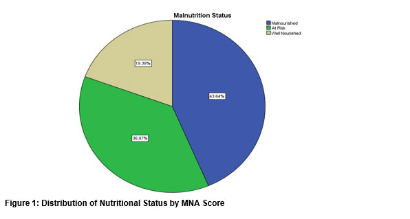
The distribution of nutritional status, as depicted in Figure 1, revealed that 144 patients (43.6%) were classified as malnourished, 122 patients (37.0%) were at risk of malnutrition, and 64 patients (19.4%) were well-nourished.
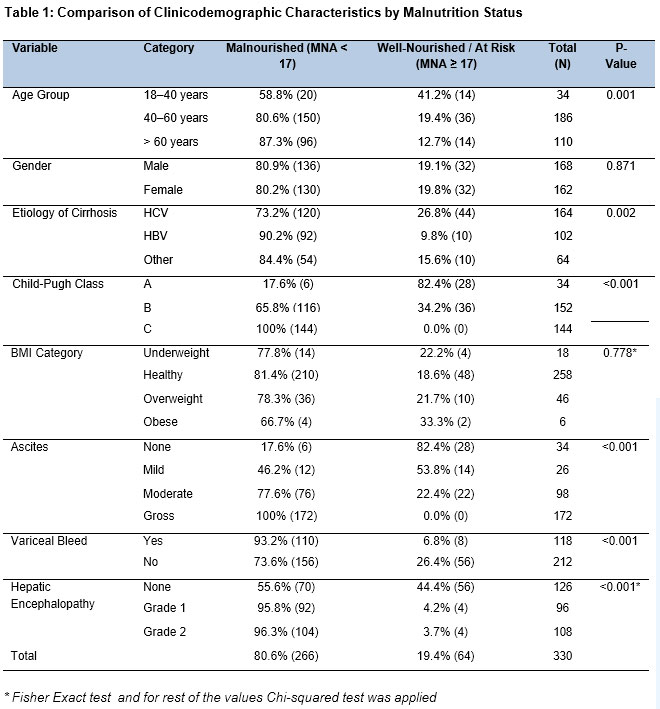
Table 1 shows significant associations between malnutrition and several clinicodemographic factors. Malnutrition was significantly more prevalent in older age groups (p=0.001). While no gender-based difference was observed, malnutrition rates varied significantly by cirrhosis etiology, being highest among HBV patients (p=0.002). A strong, direct correlation was found between worsening disease severity (higher Child-Pugh class) and malnutrition prevalence (p=0.000). Although BMI category showed no significant association, the presence and severity of ascites, a history of variceal bleeding, and the presence of hepatic encephalopathy were all significantly associated with higher rates of malnutrition (p=0.000 for each).
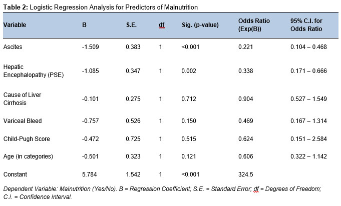
Logistic regression analysis (Table 2) identified ascites and hepatic encephalopathy (PSE) as significant independent predictors of malnutrition. The model indicates that for every one-unit increase in the severity of ascites, the odds of malnutrition decrease by a factor of 0.221 (representing a 77.3% reduction in odds), and for every one-unit increase in the grade of encephalopathy, the odds decrease by a factor of 0.338 (a 66.2% reduction). This inverse relationship in the odds ratio stems from the coding of these variables, confirming their strong association with malnutrition. Other factors, including the cause of cirrhosis, variceal bleed, Child-Pugh score, and age, did not emerge as significant independent predictors in this model 22.
Discussion
Early nutritional assessment and subsequent intervention are crucial for improving the prognosis of malnourished cirrhotic patients, potentially reducing mortality, particularly among those awaiting liver transplantation. The cornerstone of this process is a comprehensive nutritional evaluation using validated tools to assess intake and body composition, guiding appropriate dietary management 22.
In the present study, the overall prevalence of malnutrition was 80.6%, a figure comparable to the 86% reported in an Egyptian cohort by Khalil et al. 23 and consistent with findings from Naqvi et al. 24. This consistently high prevalence across diverse populations underscores the concept of cirrhosis inducing an "Accelerated starvation" state. Pathophysiologically, the cirrhotic liver's depleted glycogen stores compel the body to rely on skeletal muscle proteolysis for energy, precipitating rapid muscle wasting independent of the patient's baseline body weight 16. The mean age of our cohort (54.73 ± 11.93 years) aligns with other regional studies 1, 2. We observed a significantly heightened vulnerability to malnutrition in patients over 40 years of age. This can be attributed to the cumulative metabolic burden of chronic hepatic inflammation compounded by age-related physiological anorexia, reduced anabolic capacity, and diminished protein reserves, rendering older individuals less resilient to the catabolic effects of cirrhosis compared to younger populations 25, 26.
The mean BMI was 22.41 kg/m². While Khalil et al. reported higher BMIs 23, our findings are more consistent with those of Naqvi et al. 25. However, it is critical to acknowledge the limitations of BMI in this context. Fluid retention, such as ascites and peripheral edema, artificially inflates body weight, thereby masking the presence of underlying sarcopenia (muscle wasting). Consequently, a "Normal" BMI in a cirrhotic patient can conceal profound nutritional depletion, which is better captured by tools like the MNA or anthropometric measures like mid-arm circumference 27.
HBV was identified as the predominant etiology of cirrhosis in our study, diverging from other populations where HCV or alcohol-associated liver disease are more common 23, 28. This variation is justifiable given the regional endemicity of HBV. The particularly high malnutrition rate among HBV patients in our cohort may suggest that the chronic, decades-long inflammatory course of this infection leads to a more profound state of metabolic exhaustion compared to more acute or subacute etiologies.
Disease severity, as graded by the Child-Pugh score, emerged as the most potent predictor of nutritional status. Notably, malnutrition was universal (100%) among patients with Child-Pugh Class C cirrhosis, mirroring the findings of Tai et al. 29 and Vieira et al. 3. This relationship is explained by the progressive failure of hepatic synthetic and metabolic functions. As liver reserve deteriorates, its capacity for gluconeogenesis and protein synthesis is lost, forcing the body into a persistent catabolic state 30. This is further exacerbated by the presence of moderate-to-gross ascites (p < 0.001). Ascites contributes to malnutrition through mechanical compression of the stomach ("small stomach effect"), leading to early satiety and drastically reduced caloric intake, while repeated paracentesis results in significant loss of protein-rich fluid 31.
Finally, complications such as variceal bleeding and hepatic encephalopathy (HE) were identified as significant risk factors for malnutrition (p < 0.0001). This association is explained by a self-perpetuating "vicious cycle": HE impairs cognitive function, compromising the patient's ability to procure and consume food, while the acute catabolic stress triggered by a gastrointestinal bleed rapidly depletes already marginal nitrogen reserves 32. Logistic regression analysis confirmed that ascites and HE are the strongest independent predictors of malnutrition in our cohort, a finding consistent with the work of Saunders et al. 33 and Romeiro et al. 34. The study is limited by the fact that it was a single centered study and convenience sampling technique was used.
Conclusion
This study demonstrates that malnutrition is exceedingly common among patients with liver cirrhosis, with the highest prevalence observed in those of advanced age, with HBV-related disease, and with severe complications such as moderate-to-gross ascites and hepatic encephalopathy. The profound wasting characteristic of this population is primarily driven by the hypermetabolic state inherent to chronic liver disease, impaired nutrient assimilation, and the mechanical impediment to oral intake caused by ascites.
For clinicians, the critical takeaway is that nutritional decline represents a significant yet potentially modifiable risk factor. To effectively counteract this wasting process, the routine integration of early bedside nutritional assessment using validated tools like the MNA is imperative. We strongly advocate for a multidisciplinary management approach that includes prompt referral to dietitians for the development of personalized nutritional plans. Clinical strategies should prioritize early nutritional interventions, such as the implementation of frequent, small, protein-dense meals and late-evening carbohydrate snacks to minimize overnight fasting. The timely use of oral or enteral nutritional supplements should also be considered when standard dietary intake proves insufficient.
References
- Schuppan D, Afdhal NH. Liver cirrhosis. Lancet. 2008;371(9615):838-51. https://doi.org/10.1016/S0140-6736(08)60383-9
- Sharma B, John S. Hepatic cirrhosis. In: StatPearls [Internet]. Treasure Island (FL): StatPearls Publishing; 2022. Available from: https://www.ncbi.nlm.nih.gov/books/NBK482419/
- Kim D, Bonham CA, Konyn P, Cholankeril G, Ahmed A. Mortality trends in chronic liver disease and cirrhosis in the United States, before and during COVID-19 pandemic. Clin Gastroenterol Hepatol. 2021;19(12):2664-6.e2. https://doi.org/10.1016/j.cgh.2021.07.009
- Tsiaousi ET, Hatzitolios AI, Trygonis SK, Savopoulos CG. Malnutrition in end-stage liver disease: recommendations and nutritional support. J Gastroenterol Hepatol. 2008;23(4):527-33. https://doi.org/10.1111/j.1440-1746.2008.05369.x
- Henkel AS, Buchman AL. Nutritional support in patients with chronic liver disease. Nat Clin Pract Gastroenterol Hepatol. 2006;3(4):202-9. https://doi.org/10.1038/ncpgasthep0443
- Ney M, Li S, Vandermeer B, Gramlich L, Ismond KP, Raman M, Tandon P. Systematic review with meta-analysis: nutritional screening and assessment tools in cirrhosis. Liver Int. 2020;40(3):664-73. https://doi.org/10.1111/liv.14269
- Merli M, Riggio O, Dally L. Does malnutrition affect survival in cirrhosis? Hepatology. 1996;23(5):1041-6. https://doi.org/10.1002/hep.510230516
- Sam J, Nguyen GC. Protein-calorie malnutrition as a prognostic indicator of mortality among patients hospitalized with cirrhosis and portal hypertension. Liver Int. 2009;29(9):1396-402. https://doi.org/10.1111/j.1478-3231.2009.02053.x
- European Association for the Study of the Liver. EASL clinical practice guidelines on nutrition in chronic liver disease. J Hepatol. 2019;70(1):172-93. https://doi.org/10.1016/j.jhep.2018.06.024
- Velasco C, García E, Rodríguez V, Frías L, Garriga R, Álvarez J, et al. Comparison of four nutritional screening tools to detect nutritional risk in hospitalized patients: a multicentre study. Eur J Clin Nutr. 2010;64(9):1052-9. https://doi.org/10.1038/ejcn.2010.243
- Yasutake K, Koga S, Hokko Y, Oikawa SI, Nagata K, Tanaka T, et al. Relevance of the Mini Nutritional Assessment in cirrhotic liver disease patients. Asia Pac J Clin Nutr. 2018;27(2):300-5. https://doi.org/10.6133/apjcn.052017.04
- Praneetvatakul Y, Larpjit S, Jongcherdchootrakul K, Lertwanichwattana T. Prevalence and associated factors of malnutrition among elderly patients. J Southeast Asian Med Res. 2023;7:e0167. https://doi.org/10.55374/jseamed.v7.167
- Bauer J, Vogl T, Wicklein S, Trögner J, Mühlberg W, Sieber C. Comparison of nutritional screening tools in geriatric hospital patients. Z Gerontol Geriatr. 2005;38(5):322-7. https://doi.org/10.1007/s00391-005-0331-9
- Read JA, Crockett N, Volker DH, MacLennan P, Choy STB, Beale P, et al. Nutritional assessment in cancer. Nutr Cancer. 2005;53(1):51-6. https://doi.org/10.1207/s15327914nc5301_6
- Shin S, Jun DW, Saeed WK, Koh DH. A narrative review of malnutrition in chronic liver disease. Ann Transl Med. 2021;9(2):172. https://doi.org/10.21037/atm-20-4868
- Tandon P, Ney M, Irwin I, Ma MM, Gramlich L, Bain VG, et al. Severe muscle depletion in patients on the liver transplant wait list. Liver Transpl. 2012;18(10):1209-16. https://doi.org/10.1002/lt.23495
- Zierle-Ghosh A, Jan A. Physiology, body mass index. In: StatPearls [Internet]. 2022. Available from: https://www.ncbi.nlm.nih.gov/books/NBK535456/
- Das A, Saimala G, Reddy N, Mishra P, Giri R, Kumar A, et al. Mid-upper arm circumference as a substitute of BMI. Public Health. 2020;179:68-75. https://doi.org/10.1016/j.puhe.2019.09.010
- Yin L, Fan Y, Lin X, Zhang L, Li Y, Sun T, et al. Fat mass assessment using triceps skinfold thickness. Br J Nutr. 2022;127(10):1506-16. https://doi.org/10.1017/S0007114521002531
- Vellas B, Guigoz Y, Garry PJ, Nourhashemi F, Bennahum D, Lauque S, et al. The Mini Nutritional Assessment (MNA). Nutrition. 1999;15(2):116-22. https://doi.org/10.1016/s0899-9007(98)00171-3
- Tsoris A, Marlar CA. Use of the Child-Pugh score in liver disease. In: StatPearls [Internet]. 2022. Available from: https://pubmed.ncbi.nlm.nih.gov/31194448/
- Sapkota Y, Gnawali A, Tiwari K, Sharma TM, Thapa B. Assessment of severity of liver cirrhosis. World J Adv Res Rev. 2022;13(2):239-51. https://doi.org/10.30574/wjarr.2022.13.2.0145
- Khalil SS, Youssef MES, Mekkawy MM, Abdelmalek MO. Liver cirrhosis: nutritional status. Assiut Sci Nurs J. 2015;3(6):114-25. https://doi.org/10.21608/asnj.2015.59806
- Vieira PM, De-Souza DA, Oliveira LC. Nutritional assessment in hepatic cirrhosis. Nutr Hosp. 2013;28(5):1615-21. https://doi.org/10.3305/nh.2013.28.5.6563
- Naqvi IH, Mahmood K, Salekeen S, Akhter ST. Malnutrition and liver cirrhosis severity. Turk J Gastroenterol. 2013;24(5):415-22. https://doi.org/10.4318/tjg.2013.0637
- Cruz-Jentoft AJ, Sayer AA. Sarcopenia. Lancet. 2019;393(10191):2636-46. https://doi.org/10.1016/S0140-6736(19)31138-9
- Cederholm T, Jensen GL, Correia MITD, Gonzalez MC, Fukushima R, Higashiguchi T, et al. GLIM criteria for diagnosis of malnutrition. Clin Nutr. 2019;38(1):1-9. https://doi.org/10.1016/j.clnu.2018.08.002
- Sharif S, Abu-Dayyeh BK, Sharif S, Al-Judaibi B. Hepatitis C and malnutrition. World J Gastroenterol. 2015;21(18):5432-40. https://doi.org/10.3748/wjg.v21.i18.5432
- Tai ML, Goh KL, Mohd-Taib SH, Rampal S, Mahadeva S. Malnutrition in cirrhosis patients. Nutr J. 2010;9:27. https://doi.org/10.1186/1475-2891-9-27
- Kim HY. Nutritional intervention in liver cirrhosis. Clin Mol Hepatol. 2023;29(2):331-4. https://doi.org/10.3350/cmh.2023.0118
- Haderer M, Ebner S, Schütz B, Becker S, Geginat G, Bruns T. Malnutrition impact in cirrhosis. Nutrients. 2023;15(18):3965. https://doi.org/10.3390/nu15183965
- Chiriac S, Stanciu C, Cojocariu C, Singeap AM, Sfarti C, Cuciureanu T, et al. Sarcopenia and hepatic encephalopathy. J Clin Med. 2023;12(13):4214. https://doi.org/10.3390/jcm12134214
- Saunders J, Brian A, Wright M, Stroud M. Malnutrition in liver disease. Frontline Gastroenterol. 2010;1(2):105-11. https://doi.org/10.1136/fg.2009.000414
- Romeiro FG, Augusti L. Nutritional assessment in cirrhotic patients. World J Hepatol. 2015;7(30):2940-54. https://doi.org/10.4254/wjh.v7.i30.2940