Abstract
Background: Diabetes is an alarmingly prevalent disease in Pakistan, affecting 26.7 % of adult population according to International Diabetic Federation (IDF). Fungal sinus infections and their complications, being dominant in immunocompromised diabetic patients, need to be diagnosed timely with MRI imaging to avoid devastating complications. Objective: The main objective of our research is to prove diagnostic accuracy of MRI for the diagnosis of invasive fungal rhinosinusitis among diabetic, immuno-compromised patients.
Method: We performed an analytical cross-sectional study spanning ten months (May 2024 till February 2025) at the Radiology Department of Services Hospital, Lahore. Non-probability consecutive sampling technique was utilized through which 191 diabetic patients of age 35 to 50 years from both genders affected by invasive fungal rhinosinusitis were enrolled to constitute the study population. Each subject was examined using pre and post contrast MRI of paranasal sinuses, following which, histopathological confirmation was made and the results were analyzed using SPSS version 25.0 software to measure diagnostic accuracy of MRI taking histopathology as gold standard.
Results: Among the 191 diabetic patients, most of them fell in the age range of 40 to 50 years (72%) while the rest of them were aged 35 to 39 years (28%). The mean age was 42.76 years ± 5.37 years. Pre contrast MRI accompanied by Post contrast MRI has Sensitivity: 84.37%, Specificity: 88.05 %, PPV: 58.69%, NPV: 96.55 %, along with overall accuracy: 87.43
% to detect invasive fungal rhinosinusitis.
Conclusion: Magnetic resonance imaging has substantial sensitivity and specificity for diagnosing Invasive Fungal Rhinosinusitis with high degree of confidence, furthermore it pinpoints localization of extra sinus fungal invasion to provide a roadmap for surgical planning thus evading critical consequences.
Keywords: Invasive, Rhinosinusitis, Diabetics, Histopathology.
Introduction
Invasive fungal rhinosinusitis (IFRS) is an aggressive fungal disease that involves the nose and paranasal sinuses, further spreading quickly through sinus walls into adjacent structures including the orbit, bones, vessels, and intracranial soft tissues such as the brain parenchyma, resulting in detrimental effects 1, 2. This infection is highly prevalent in diabetic patients due to their immunocompromised status 3, 4. In developing countries like Pakistan, there has been a notable surge in IFRS cases over the last two to three decades. Poor glycemic control, diabetic ketoacidosis due to undiagnosed diabetes, diabetic neuropathy, oxidative stress, and diabetic vasculopathy are major risk factors for fungal colonization.
Magnetic Resonance Imaging (MRI) with pre-contrast and post-contrast images provides excellent soft tissue resolution and has potential for early detection of intra-orbital and intracranial fungal extension, thereby reducing morbidity and mortality 5. MRI is also a useful tool to monitor disease progression and guide treatment strategies. Crucial MRI features for identifying extra-nasal, intra-orbital, and intracranial extensions include similar signal intensity mass lesions in the pterygomaxillary space, replacement of normal extraconal fat signals, involvement of the nasolacrimal drainage apparatus, and brain parenchyma involvement 6. These findings may present clinically with visual disturbances and neurological problems 7.
As IFRS involves vessels, MRI can reveal compression, loss of normal signal voids, pseudoaneurysm formation, and intraluminal filling defects in the internal carotid artery and superior ophthalmic vein, with or without cavernous sinus thrombosis 8, 9. Other lethal complications of fungal rhinosinusitis seen on MRI include orbital/periorbital infections, meningitis, intracerebral abscess, and venous sinus thrombosis. MRI excels in identifying extension to perineural regions and cavernous sinuses, both severe consequences of fungal sinus disease. Fulminant fungal invasion leads to visual disturbances and high mortality due to the presence of these findings 10. Delayed diagnosis of osseous involvement contributes to documented mortality rates of 50–80% 1. Thus, MRI is a crucial diagnostic modality that helps clinicians plan medical management or interventional strategies, including endoscopic surgery or aggressive surgical debridement 11.
Histopathology is employed to verify IFRS and to identify the causative fungal strain 12. Factors impacting MRI diagnostic accuracy in IFRS among diabetic patients include timing of the scan, image resolution, expertise of the reporting team, and comparison with histopathology. A recent local study in Karachi reported MRI sensitivity of 82.6%, specificity of 89.4%, diagnostic accuracy of 88.3%, PPV of 60.3%, and NPV of 96.3%, taking histopathology as the gold standard 1.
Since comparable local literature is limited, more research is required as many previous studies were conducted in European countries with different demographics. Our study focused on assessing the diagnostic performance of pre- and post-contrast MRI in invasive fungal rhinosinusitis among diabetic patients in the local population, with histopathology taken as the reference gold standard.
Method
This cross-sectional analysis was carried out at the Radiology Department of Services Hospital Lahore over ten months (May 2024 to February 2025) with permission from the institutional review board (IRB approval letter Ref No. IRB/2024/1321/SIMS dated 25-04-24). To calculate the sample size of 191, prevalence was considered as 17.8%, precision value as 58.69%, with expected sensitivity and specificity of 82% and 89%, respectively. Non-probability consecutive sampling technique was adopted to include 191 patients, with a 95% confidence level and 5% margin of error.
Diabetic participants referred with clinical suspicion of IFRS for imaging evaluation provided written informed consent. The included subjects comprised diabetic patients of both genders, aged 35–50 years, with clinical suspicion of invasive fungal rhinosinusitis and bony erosion on CT. Patients previously documented with primary cerebral or orbital infections, paranasal sinus tumors, cases unfit for surgery, non-diabetics, those allergic to gadolinium contrast, and those contraindicated for MRI due to metallic prosthesis were excluded.
All patients underwent cranial MRI including nose and sinus cavities using a 1.5 Tesla MRI machine (Canon Japan, Vantage-Titan model). Pre-contrast images (T1WI, T2WI, T2 FAT SAT) and post-contrast multiplanar images using intravenous gadolinium contrast agent were obtained. All scans were labeled positive or negative depending on MRI findings of invasive fungal rhinosinusitis after evaluation by an expert radiologist. These findings were compared with histopathological results, taken as the gold standard.
A preconceived proforma was used to assemble the data. The gathered data was analyzed using Statistical Package for Social Sciences (SPSS) version 25. Qualitative variables, including diagnosis of IFRS using MRI and histopathology, were expressed as frequencies and percentages. Quantitative variables such as age and BMI were expressed as mean ± standard deviation.
Results
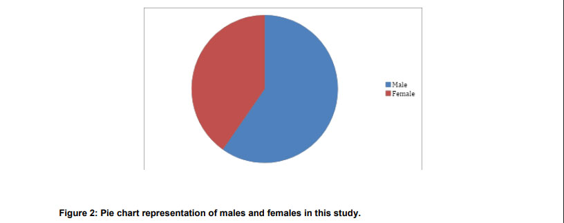
Out of 191 patients, the mean age was calculated as 42.76 ± 5.37 years, with an age range of 35–50 years. The gender distribution among the participants included 114 males (59.7%) and 77 females (40.3%). The mean BMI was 27.06 ± 3.60 kg/m², with minimum and maximum values of 23.46 and 30.66 kg/m², respectively.
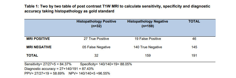
We calculated the diagnostic accuracy of pre-contrast MRI accompanied by post-contrast MRI for invasive fungal rhinosinusitis (IFRS), along with predictive values, using histopathology as the gold standard. The analysis provided sensitivity, specificity, positive predictive value (PPV), negative predictive value (NPV), and overall diagnostic accuracy for MRI in detecting IFRS among diabetic patients.
Discussion
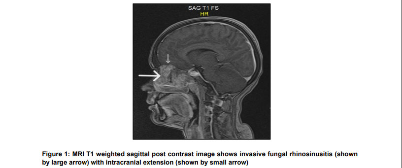
Invasive fungal rhinosinusitis (IFRS) poses a significant life-threatening risk to diabetic immunocompromised individuals due to devastating intracranial and intra-orbital complications, triggering high mortality. Early diagnosis using a multidisciplinary approach is imperative for swift intervention 4. Although widely used, CT scan often underestimates the full extent of IFRS, with its primary utility limited to visualizing sinus opacification and bony erosions. Histopathology, considered the gold standard for diagnosing IFRS, remains invasive as it requires tissue sampling. This study underscores the enhanced diagnostic capability of MRI in identifying IFRS extension beyond the sinuses into adjacent soft tissues, orbit, and brain. MRI demonstrated high sensitivity and specificity, making it the most reliable and accepted diagnostic investigation. Its excellent soft tissue contrast, enhanced spatial resolution, and noninvasive radiation-free properties reinforce its clinical utility. Fungal hyphae with paramagnetic properties appear hypointense on both T1 and T2 weighted images, indicating invasion of surrounding structures (Fig-I). Nevertheless, signal heterogeneity can arise from fluid and proteinaceous secretions within the paranasal sinuses 13.
In our study, MRI exhibited sensitivity of 84.37%, specificity of 88.05%, and diagnostic accuracy of 87.43% for diagnosing IFRS in diabetics. This demonstrates MRI’s proficiency in recognizing true positives and true negatives. Hassan F et al. reported similar values: sensitivity 82.6%, specificity 89.4%, and diagnostic accuracy 88.3%. Abokoura et al. found MRI to have 91.7% sensitivity and 100% specificity for detecting IFRS complications 12. Patients in our study had a mean age of 42.76 ± 5.37 years, differing from Abokoura’s cohort (54.67 ± 5.26 years), likely due to the younger immunocompromised population in our country, consequent to high diabetes prevalence, poor socioeconomic status, and demographic differences.
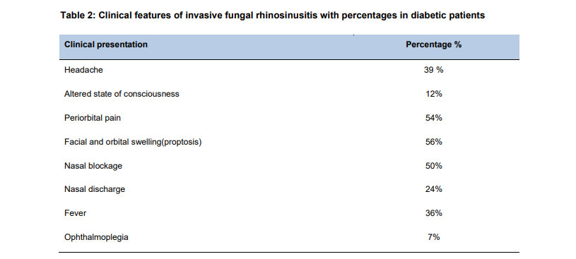
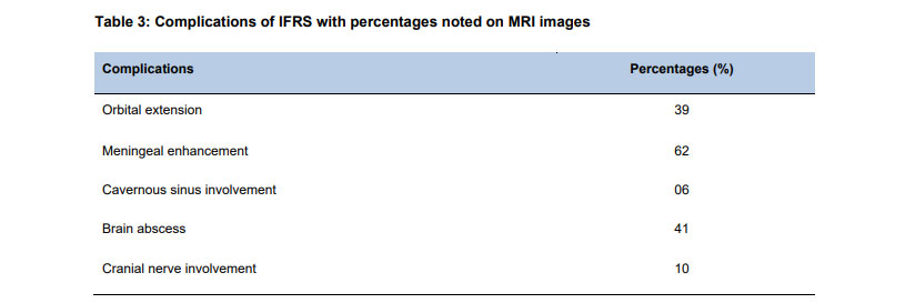
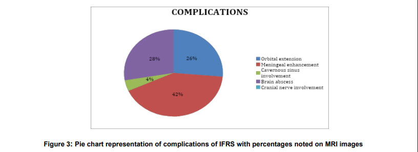
Our findings on the most common clinical feature of facial and orbital swelling (proptosis) in 56% of patients are consistent with Qazi ZU et al. 13. Cho et al. also reported MRI’s capability for extra-sinus invasion with orbital involvement (75%), pterygopalatine fossa (75%), masticator space (50%), and skull base invasion (75%) 14. Serris A et al. described two routes of fungal spread to the brain: direct extension from ear or paranasal sinuses, and blood-borne spread. The latter leads to multiple brain abscesses, while direct extension results in meningitis and cranial nerve palsies 15. Diffusion-weighted and contrast-enhanced MRI sequences can diagnose optic nerve ischemia and myositis, as discussed by Petroulia VD et al. 16 and Lever M et al. 17. Lersy F et al. noted that perineural spread is uncommon in immunocompetent patients and in aspergillosis cases 18.
Bhatia H et al. highlighted that coronal MRI imaging confirms direct or indirect extension into the cavernous sinus and resultant thrombosis, seen as enlargement with absent internal flow voids and diffusion restriction 19. Our study emphasizes MRI’s pivotal role in guiding IFRS treatment. Patterson TF et al. concluded that optimal IFRS management incorporates medical and surgical intervention, including antifungal therapy and debridement. Surgical debridement removes necrotic tissue, improves sinus drainage, and reduces recurrence risk 20.
Conclusion
MRI is indicated for early diagnosis of IFRS to reduce mortality and avoid its life-threatening complications like intracranial and intra-orbital extension with resultant thrombosis/ blockage of cavernous sinus. Our study endorses that plain and post contrast MRI sequences are best to evaluate invasive fungal rhinosinusitis in early phase of disease and to avoid extensive complications
References
- Hassan F, Mansoor A, Iqbal J, et al. Diagnostic accuracy of MRI in detection of invasive fungal sinusitis taking histopathology as a gold standard. Pakistan Journal of Radiology. 2021;31(3):171-176. Available from: https://www.researchgate.net/publication/359061714_diagnostic_accuracy_of_mri_in_detection_of_invasive_fungal_sinusitis_taking_histopathology_as_a_gold_standard
- Erami M, Aboutalebian S, Hezaveh SJH, Ghazvini RD, Momen-Heravi M, Jafari Y, et al. Microbial and clinical epidemiology of invasive fungal rhinosinusitis in hospitalized COVID-19 patients, the divergent causative agents. Medical Mycology. 2023;61(3):myad020. https://doi.org/10.1093/mmy/myad020
- Taghian E, Abtahi SH, Mohammadi A, Hashemi SM, Ahmadikia K, Dolatabadi S, Mohammadi R. A study on the fungal rhinosinusitis: Causative agents, symptoms, and predisposing factors. Journal of Research in Medical Sciences. 2023;28:12. https://doi.org/10.4103/jrms.jrms_270_22
- Yağmur AR, Çufalı ÖF, Çolak M, et al. Invasive fungal rhinosinusitis, clinical manifestations, and prognostic values: a case series audit. Egyptian Journal of Otolaryngology. 2023;39:189. https://doi.org/10.1186/s43163-023-00551-8
- Lai J, Wang B, Petrik M, Beziere N, Hammoud DA. Radiotracer development for fungal-specific imaging: past, present, and future. Journal of Infectious Diseases. 2023;228(4):259–269. https://doi.org/10.1093/infdis/jiad067
- Kondapavuluri SK, Anchala VKR, Bandlapalli S, et al. Spectrum of MR imaging findings of sinonasal mucormycosis in post COVID-19 patients. British Journal of Radiology. 2021;94(1127):20210648. https://doi.org/10.1259/bjr.20210648
- Huang YF, Liang KL, Liang CY, Yang PC, Chen JP, Wei LC. Acute invasive fungal rhinosinusitis-related orbital infection: a single medical center experience. Journal of Ophthalmology. 2021;2021:9987871. https://doi.org/10.1155/2021/9987871
- Idowu OO, Karl A, Soderlund KA, et al. Magnetic resonance imaging prognostic findings for visual and mortality outcomes in acute invasive fungal rhinosinusitis. American Journal of Ophthalmology. 2022;129(11):1313–1322. https://doi.org/10.1016/j.amjophth.2022.06.017
- Chen A, Pietris J, Bacchi S, et al. Imaging features of invasive fungal rhinosinusitis: a systematic review. Canadian Association of Radiologists Journal. 2024;75(3):601–608. https://doi.org/10.1177/08465371241227424
- Kovanda LL, Marty FM, Maertens J, et al. Impact of mucositis on absorption and systemic drug exposure of isavuconazole. Antimicrobial Agents and Chemotherapy. 2017;61(6):e00101-17. https://doi.org/10.1128/AAC.00101-17
- Roland LT, Humphreys IM, Le CH, et al. Diagnosis, prognosticators, and management of acute invasive fungal rhinosinusitis: multidisciplinary consensus statement. International Forum of Allergy & Rhinology. 2023;13(9):1615–1714. https://doi.org/10.1002/alr.23132
- Abokoura S, Youssef A, Nada A, et al. MRI manifestations of vascular complications in acute invasive fungal rhinosinusitis. Egyptian Journal of Radiology and Nuclear Medicine. 2023;54:133. https://doi.org/10.1186/s43055-023-01081-2
- Qazi ZU, Latif S, Awan SM. Orbital involvement in sinonasal diseases. Journal of Ayub Medical College Abbottabad. 2016;28(4):687–693. Available from: https://www.ayubmed.edu.pk/jamc/index.php/jamc/article/view/1189
- Cho SJ, Choi YJ, Cho KJ, et al. Imaging findings in chronic invasive fungal infection of paranasal sinuses. Journal of Neuroradiology. 2021;48(5):325–330. https://doi.org/10.1016/j.neurad.2021.02.005
- Serris A, Benzakoun J, Danion F, et al. Cerebral aspergillosis in the era of new antifungals: CEREALS study. Journal of Infection. 2022;84(2):227–236. https://doi.org/10.1016/j.jinf.2021.11.014
- Petroulia VD, Brügger D, Hoepner R, et al. MRI signs in optic neuropathy differentiation. British Journal of Ophthalmology. 2023;107:121–126. https://doi.org/10.1136/bjophthalmol-2021-319537
- Lever M, Wilde B, Pförtner R, et al. Orbital aspergillosis: case report and review. BMC Ophthalmology. 2021;21(1):22. https://doi.org/10.1186/s12886-020-01773-7
- Lersy F, Royer-Leblond J, Lhermitte B, et al. Cerebral mucormycosis: neuroimaging findings and histopathological correlation. Journal of Neurology. 2022;269:1386–1395. https://doi.org/10.1007/s00415-021-10701-8
- Bhatia H, Kaur R, Bedi R. MR imaging of cavernous sinus thrombosis. European Journal of Radiology Open. 2020;7:100226. https://doi.org/10.1016/j.ejro.2020.100226
- Patterson TF, Thompson GR, Denning DW, et al. Practice guidelines for the diagnosis and management of aspergillosis. Clinical Infectious Diseases. 2016;63(4):e1–e60. https://doi.org/10.1093/cid/ciw326